
BMEssentia Platform / Device library / Intravascular Ultrasound (IVUS) System
Device library
Intravascular Ultrasound (IVUS) System
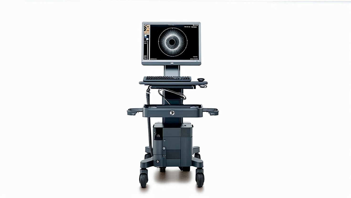
Intravascular Ultrasound (IVUS), introduced in the late 1980s, uses a tiny ultrasound probe on a catheter to image blood vessels from the inside, revealing plaque buildup invisible to traditional X-ray angiography. It provides a 360-degree view of artery walls, helping cardiologists assess plaque type, guide stent placement, and reduce complications like stent thrombosis. Modern IVUS systems, like those from Philips and Boston Scientific, offer real-time imaging without radiation, improving outcomes in heart and peripheral artery procedures.
The 13 sections, in brief
Short summaries of each section. The full lessons, images and quizzes are in the Platform.
Overview
A cardiologist is looking at an angiogram of a coronary artery and the narrowing is about forty percent. Borderline. Not obviously significant, not obviously safe.
Operations
Follow the catheter. A guidewire is already across the lesion from the angiographic procedure. The IVUS catheter is advanced over that wire until its transducer sits distal to the segment of interest. Then it is pulled back, usually by a motorised device at a constant speed, while the transducer images continuously.
Importance
Three arguments, and the third is the one that changed practice. First, it sees the disease rather than its shadow. Plaque burden, remodelling, calcification depth, and the true reference vessel size. An artery that looks mildly diseased on angiography is frequently severely diseased on ultrasound, and the reverse happens too.
Physics
Physics Involved in the Intravascular Ultrasound (IVUS) System: Concept 1, Catheter-Based Acoustic Imaging and Lumen Reconstruction At the heart of IVUS lies a concept both breathtaking and delicate, seeing the artery from within.
Features
Why the frequency is so high, and what it costs The trade in ultrasound is always the same. Higher frequency gives finer resolution and travels less far. An abdominal probe runs low because it has to reach twenty centimetres. This transducer has to reach about five millimetres.
Applications
Optimising stent deployment, the application with the evidence The largest use and the one supported by randomised trials rather than by plausibility. Vessel sizing from the true outer dimension, not from the contrast silhouette. Which consistently produces larger stent choices than angiography suggests, and larger well-expanded stents stay open.
Physiology
Three layers, and the device sees all three. The inner lining, a single cell layer thick, which is the active surface. It is anticoagulant, it controls vessel tone, and it regulates what crosses into the wall. Atherosclerosis begins when it stops doing those things properly.
Safety
The complication rate for this device is low, and the specific hazard follows directly from that. It is an additional catheter, inserted into a coronary artery, for information rather than for treatment.
Advancements
Forget release years. This device has a shape that is worth naming precisely, because it is unusual. The technology matured long before the evidence arrived, and the evidence arrived long before the adoption did. Somebody put a transducer on a catheter and discovered the artery wall, which angiography had never shown.
In Research
Almost every device in this library has the same problem: it was adopted on plausibility and never properly tested. It was tested. Randomised trials showed that imaging-guided stenting reduces target lesion failure. The evidence exists, it is reasonably strong, and the device is still used in a minority of procedures in most health systems.
Logistics
An engineer who understands this device is useful. An engineer who understands that the console is frequently given away and the catheter is the entire business understands what is actually being sold here. Because this is the cleanest razor-and-blade structure in the library.
Alternatives
An alternative is not a competing product. It is another way of answering the clinical question, and this device faces two completely different competitors because it is asked two completely different questions. When the question is what does this artery look like, the competitor is optical imaging and the comparison is about resolution against penetration.
Ethical
This device does not raise the questions most of this library raises. Nobody is unconscious for weeks, nobody is being irradiated systemically, nobody is having their consciousness taken from them. The ethical problem here is narrower and, in its way, more uncomfortable.
Previous: Intraoperative Neurophysiological Monitoring (IONM) System. Next: Laryngoscope. All 69 devices.
Learn it properly, inside the Platform
22 courses, the 69-device library with 13 sections per device, BMEssentials and BMEssence guides and MedTech AI. Start with a 7-day free trial, no card.
Start the 7-day free trial