
BMEssentia Platform / Device library / Vitrectomy Machine
Device library
Vitrectomy Machine
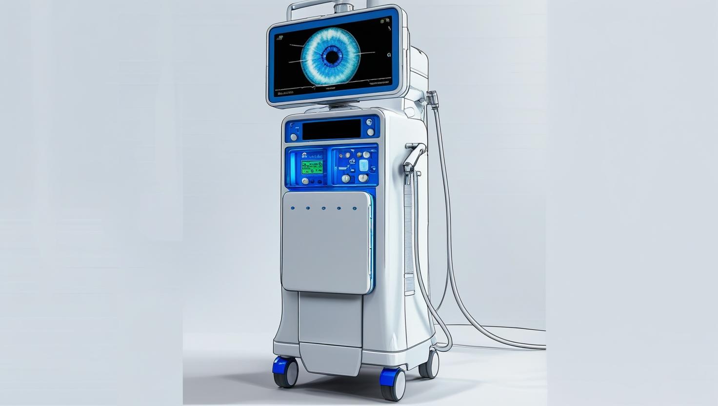
A Vitrectomy Machine is used in delicate eye surgeries to remove the vitreous gel from the eye's interior. This is often necessary to treat conditions like retinal detachment, eye infections, or bleeding inside the eye. The machine precisely cuts and suctions the gel while maintaining the eye's pressure and stability. Modern vitrectomy systems are so advanced that they allow surgeons to perform microsurgery through tiny incisions-often without stitches!
The 13 sections, in brief
Short summaries of each section. The full lessons, images and quizzes are in the Platform.
Overview
A surgeon is about to put a cutting instrument inside somebody's eye, and there are exactly two places on the whole eyeball where that is survivable. Go in at the front and you destroy the lens. Go in at the back and you cut through the retina, which is the thing you are there to save.
Operations
Follow the eye, then follow the fluid, because the fluid balance is the device. Start with the ports. Three small incisions are made through the white of the eye, in the safe ring behind the cornea. In modern systems a trocar is used to place a cannula, which is a short tube that stays in the sclera for the whole operation and gives instruments a door to pass through.
Importance
The first argument is about what this machine prevents, and it is absolute. A detached retina goes blind. Not slowly, and not partially. The retina is fed from behind by the choroid, and separating the two starves the photoreceptors. They begin to die within days and the damage becomes permanent.
Physics
Physics Involved in the Vitrectomy machine: Concept 1, Core System Architecture and Closed-Loop Vitreous Removal Mechanism A vitrectomy machine is not just an aspiration pump-it is a microfluidic surgical ecosystem. Its role is to remove the vitreous humor, a gel-like matrix composed of collagen fibrils and hyaluronic acid-from the posterior chamber of the eye without destabilizing intraocular pressure (IOP).
Features
Start outside the machine entirely, because the anatomy dictates the design. The eye is built in layers, and each one is a hazard. In front is the lens, a transparent structure that has no blood supply and no capacity to repair. Touch it and it goes cloudy, permanently.
Applications
The core emergency application and the operation that defines the specialty. The vitreous gel shrinks and separates from the retina with age, which is normal and happens to most people. Where it is stuck firmly, that separation tears a hole. Fluid passes through the hole, collects underneath the retina, and lifts it off the layer that supplies it with blood and nutrients.
Physiology
This is the section where the eye earns its reputation as the most elegant organ in the body. Everything this machine does is a response to four facts: what the vitreous is, how the retina is held on, how the retina is fed, and why the eye has no ability to repair any of it.
Safety
The defining safety fact about this device is the size of the margin. The retina is about a quarter of a millimetre thick. The instruments are held by hand, inside a sphere two centimetres across, viewed through a microscope at high magnification. A movement of half a millimetre in the wrong direction is the difference between removing a membrane and tearing the retina under it.
Advancements
Forget release years. This device has one of the clearest improvement stories in the library: it started as a large single instrument that only the boldest surgeons would use, and became a precise multi-port system that made an entire subspecialty possible.
In Research
Retinal detachment surgery works. The retina goes back, the break is sealed, the eye looks good at two weeks. And in a meaningful proportion of cases the patient's own healing response then pulls it off again. Cells escape through the retinal break, change character, grow on the retinal surfaces and contract.
Logistics
An engineer who understands this machine is useful. An engineer who understands that the console is a loss leader and the pack is the business understands what is actually being sold. A vitrectomy platform is a substantial capital purchase, and it is not where the money is.
Alternatives
An alternative is not a competing product. It is another way of answering the clinical question, and here the questions are separate. Can this retina be reattached without entering the eye? Can this disease be stopped before it needs surgery at all? The second question has done more for vision in the last generation than any improvement to the machine.
Ethical
Blindness is not like other disability. It is the outcome people fear most after death, consistently, in every survey that asks. Which changes the dynamics of every conversation in this device. A patient who has been told they might lose their sight will agree to almost anything. They are not weighing options. They are frightened, and the person offering the operation knows it.
Previous: Vagus Nerve Stimulator (VNS). All 69 devices.
Learn it properly, inside the Platform
22 courses, the 69-device library with 13 sections per device, BMEssentials and BMEssence guides and MedTech AI. Start with a 7-day free trial, no card.
Start the 7-day free trial