
BMEssentia Platform / Device library / Computed Tomography
Device library
Computed Tomography
Computed Tomography (CT), introduced in 1972 by Godfrey Hounsfield, revolutionized imaging by combining X-rays with computer processing to create detailed 3D images of organs, bones, and tissues. Early CT scanners took minutes per scan, exposing patients to high radiation, but modern systems, like Siemens' SOMATOM, produce high-resolution images in seconds with reduced doses. Advanced CTs use AI to enhance image clarity and detect abnormalities like tumors or brain bleeds, making them critical for diagnostics in emergencies and oncology.
The 13 sections, in brief
Short summaries of each section. The full lessons, images and quizzes are in the Platform.
Overview
It is twenty past three in the morning and a man has been brought in from a road accident with a blood pressure that will not hold. He is unconscious, he is bleeding somewhere, and nobody knows where. His abdomen is rigid but abdomens lie. His chest sounds equal but chests lie too.
Operations
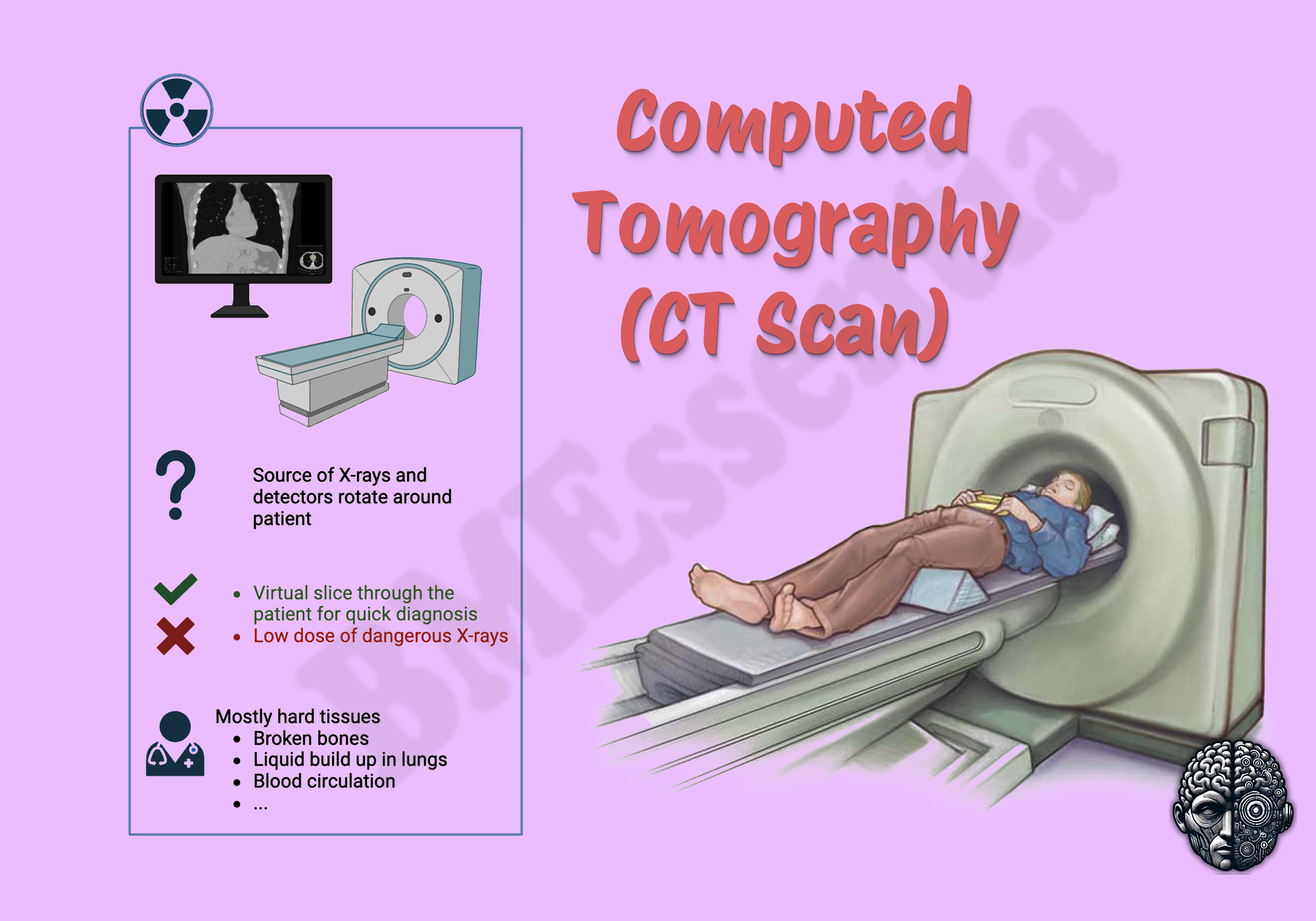
Follow a scan and the machine explains itself. The patient lies on a table that moves through a short ring. Inside that ring, mounted on a rotating gantry, an X-ray tube fires a fan of radiation through the body, and directly opposite it an arc of detectors measures how much got through along every path.
Importance
The case for CT is speed and completeness together, and no other cross-sectional modality offers both. A whole body in seconds, in a patient who is unconscious, ventilated, bleeding, and surrounded by monitoring equipment, none of which has to be removed. That single capability reorganised emergency medicine.
Physics
At the heart of every CT scanner lies a source of invisible yet precisely engineered energy, the X-ray beam. " It is a meticulously crafted output of electron-photon interactions inside a high-voltage vacuum tube designed to deliver diagnostic information at the smallest possible biological cost.
Features
The X-ray tube, and the component that limits everything Almost every performance limit in a CT scanner traces back to one component that is doing something brutal. Inside the tube, electrons are accelerated across a high voltage and slammed into a metal target. A small fraction of that energy becomes X-rays. The overwhelming majority becomes heat. Not a little heat.
Applications
Emergency medicine, which this machine reorganised Trauma. The whole-body scan in a major trauma patient has become standard in systems that can support it, and the reason is simple: a patient with multiple injuries cannot reliably tell you what hurts, examination is unreliable in the unconscious, and injuries missed early are injuries found late.
Physiology
Here is the difficulty with CT physiology, and it is worth naming before we start. This machine does not measure a biological process. It measures how strongly tissue absorbs X-rays, which is a physical property, not a physiological one. So there is a temptation to say there is no physiology to teach.
Safety
Every other safety section in this library is about a hazard you can point at. A magnet that never switches off. A patient who walks out of the department radioactive. Four metal arms inside an abdomen. CT has none of that. The source is inside the machine. The room is safe when it is not scanning. Nothing is left behind in the patient. Nothing follows them home.
Advancements
Forget release years. They rot, and they teach you nothing. What you want is the shape of how this field moved, because the shape predicts direction and stays true for a decade. The mathematics existed before the machine. The problem of reconstructing an object from its projections was solved on paper long before anyone considered pointing X-rays at a person.
In Research
CT is the most quantitative imaging modality in routine use and one of the most performed. Its physics is completely understood. Its mathematics was solved before the machine existed. There is nothing mysterious about how it works. Nobody can tell you what fraction of the CT scans performed in any health system actually changed a patient's management.
Logistics
An engineer who understands how a CT scanner works is useful. An engineer who understands how it arrives, gets licensed, gets a room built around it, gets installed, and then gets its tube replaced three times over a decade runs a department or a distributorship.
Alternatives
An alternative is not a competing product. It is another way of answering the clinical question, and the honest comparison changes completely depending on what is being asked, about whom, and in what circumstances. CT is the default cross-sectional modality in most of the world, which changes the shape of this section entirely.
Ethical
CT raises an ethical problem that almost no other device has, and it is worth stating precisely before anything else. The risk from any single scan is small enough to be invisible. Which means every individual decision to scan looks harmless. And the aggregate does not.
Previous: Colonoscope. Next: Conventional X-ray Machine. All 69 devices.
Learn it properly, inside the Platform
22 courses, the 69-device library with 13 sections per device, BMEssentials and BMEssence guides and MedTech AI. Start with a 7-day free trial, no card.
Start the 7-day free trial