
BMEssentia Platform / Device library / Conventional X-ray Machine
Device library
Conventional X-ray Machine
Conventional X-ray machines, discovered by Wilhelm Röntgen in 1895, revolutionized medicine by producing images of bones and tissues using radiation, earning him the first Nobel Prize in Physics. Early machines were slow and exposed patients to high radiation, but modern systems use digital detectors for sharper images with lower doses. Today's X-ray technology, like portable units from Canon, can deliver results in seconds and integrate with AI for faster diagnosis of fractures or lung conditions, making them indispensable in medical imaging.
The 13 sections, in brief
Short summaries of each section. The full lessons, images and quizzes are in the Platform.
Overview
Full Name of the Device: Conventional X-ray Machine (Radiography Machine, Standard X-ray) Alternate Names (if any): Plain Film X-ray, Roentgenogram (historical term), Radiographic X-ray, Digital Radiography Conventional X-ray Machine: Uses ionizing radiation to capture internal images, crucial for assessing bones and certain soft tissues quickly and efficiently.
Operations
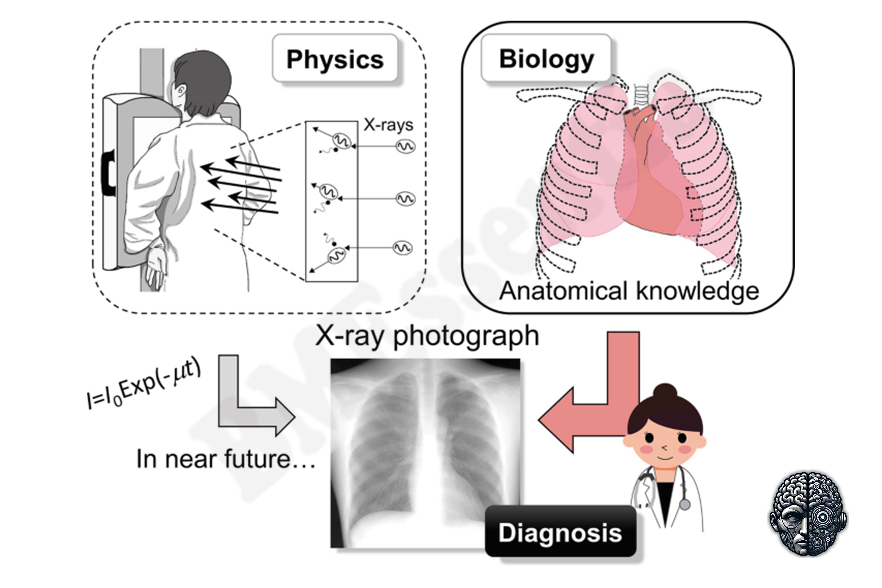
The X-ray machine operates by generating ionizing radiation through the interaction of high-speed electrons with a metal target. The machine emits X-rays in a focused beam, which passes through the body and is absorbed or scattered by tissues based on their density. Denser materials, like bone, absorb more X-rays and thus appear white on the resulting image.
Importance
X-ray machines are critical diagnostic tools due to their ability to produce fast, non-invasive images of bones and internal structures. They are the most commonly used imaging technique for bone fractures, lung conditions, and dental issues, and they serve as the first-line diagnostic tool in trauma care.
Physics
Physics Involved in the Conventional X-ray Machine: Concept 1, The X-Ray Generation Chain: Cathode-Anode Dynamics and Bremsstrahlung Production in Conventional Radiography The heart of a conventional X-ray system is a vacuum tube that converts electrical energy into high-frequency electromagnetic radiation by decelerating electrons.
Features
One of the most significant features of conventional X-ray machines is their ability to provide quick and high-resolution imaging of bones, making them indispensable in emergency rooms and trauma care settings. Their versatility allows them to be used for a wide range of applications, including chest X-rays, dental imaging, abdominal imaging, and mammography.
Applications
Working Premise and Procedure (Step-by-Step) Patient preparation for an X-ray is straightforward and typically involves removing any metallic objects, such as jewelry, belts, or eyeglasses, that could interfere with the image. Patients are also asked to wear a hospital gown if the area being examined is not easily accessible.
Physiology
X-ray imaging is used to assess various body systems, each requiring different techniques based on the density and composition of the tissues being imaged: Skeletal System: X-rays are the gold standard for imaging the bones and joints. They are used to detect fractures, bone dislocations, degenerative bone diseases, and joint abnormalities like arthritis.
Safety
X-rays use ionizing radiation, which can pose risks if exposure is not carefully controlled. While the radiation doses used in diagnostic X-rays are relatively low, repeated or prolonged exposure increases the risk of radiation-induced cancers over time.
Advancements
Recent Advances and Future Trends in X-ray Technology One of the most significant advancements in X-ray technology is the widespread adoption of digital radiography (DR), which replaces traditional film-based systems. Faster Image Acquisition: Digital systems allow for near-instantaneous image capture, reducing the time patients spend waiting for results.
In Research
X-ray imaging is superior for visualizing bones and detecting fractures or dislocations, while MRI excels at providing detailed images of soft tissues, such as muscles, tendons, and the brain. MRI is often preferred when soft tissue injuries or conditions like ligament tears or brain tumors are suspected, as it offers greater contrast between different tissue types.
Logistics
Conventional X-ray machines are relatively affordable compared to advanced imaging modalities like MRI or CT. A standard X-ray machine can range in cost from $50,000 to $200,000, depending on the system's capabilities and whether it includes features like digital radiography or fluoroscopy. Portable X-ray machines are more affordable, typically costing between $30,000 and $100,000.
Alternatives
MRI is often used as an alternative to X-ray when soft tissue imaging is required. It is ideal for diagnosing conditions like brain tumors, ligament injuries, and spinal cord issues. MRI does not involve ionizing radiation, making it safer for certain populations, but it is more expensive and time-consuming than X-ray imaging.
Ethical
Ethical Considerations and Future Directions One of the primary ethical considerations surrounding X-ray imaging is the risk of radiation exposure. Although modern X-ray machines use lower doses of radiation, repeated exposure, especially in vulnerable populations like children and pregnant women, raises concerns about long-term health risks.
Previous: Computed Tomography. Next: Cranial Drill System. All 69 devices.
Learn it properly, inside the Platform
22 courses, the 69-device library with 13 sections per device, BMEssentials and BMEssence guides and MedTech AI. Start with a 7-day free trial, no card.
Start the 7-day free trial