
BMEssentia Platform / Device library / Dermatoscope
Device library
Dermatoscope
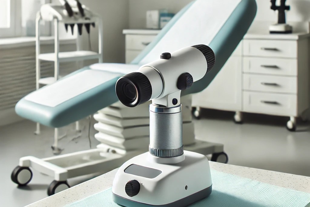
The dermatoscope is a handheld diagnostic tool that magnifies and illuminates skin lesions using polarized light or fluid immersion, enabling early detection of melanoma and other skin cancers. Modern versions feature high-resolution imaging, AI-assisted analysis, and digital tracking of suspicious moles over time. This non-invasive device improves diagnostic accuracy by 20-30% compared to naked-eye exams. Advanced models integrate with telemedicine platforms, allowing remote specialist consultations. Essential in dermatology clinics worldwide, it transforms skin exams into precise, potentially life-saving evaluations through enhanced visualization of subsurface structures. Portable.
The 13 sections, in brief
Short summaries of each section. The full lessons, images and quizzes are in the Platform.
Overview
Alternate Names: Skin surface microscope, Epiluminescence microscope, Skin scanner Dermatoscopy/Dermoscopy, The examination of skin lesions with a dermatoscope to assess structures and patterns that aren't visible to the naked eye. Polarized Light, A lighting technique used in modern dermatoscopes to reduce surface reflection, allowing clearer visualization of subsurface structures without needing immersion fluids.
Operations
The dermatoscope functions by magnifying the skin and using polarized or non-polarized light to eliminate surface reflection, enabling the visualization of subsurface skin structures. The dermatoscope is a magnifying optical instrument that typically provides 10x magnification of the skin.
Importance
The dermatoscope has revolutionized the field of dermatology by enhancing the ability of clinicians to detect and diagnose skin cancer and other skin conditions early and accurately. Early Detection of Skin Cancer: One of the primary reasons for the widespread adoption of dermatoscopes in clinical practice is their role in the early detection of skin cancer, particularly melanoma.
Physics
Physics Involved in the Dermatoscope Device: Concept 1, Polarized Illumination and Subsurface Visualization in Dermatoscopy The dermatoscope is a handheld optical instrument designed to reveal the micro-architecture of human skin by controlling how light enters, interacts, and returns from its layers.
Features
The dermatoscope is a highly specialized device with several unique features that make it essential for dermatological practice. Polarized vs. Non-Polarized Light: One of the most significant advancements in modern dermatoscopes is the use of polarized light. Polarization eliminates the need for immersion fluids by reducing surface reflection and glare, allowing clinicians to see deeper into the skin.
Applications
Working Premise and Procedure (Step-by-Step) The dermatoscope operates through a structured process that includes patient preparation, device operation, and the interpretation of images. Before starting the examination, the patient is informed about the procedure and the purpose of the dermatoscopy.
Physiology
The dermatoscope allows for the detailed examination of various skin structures that are essential for diagnosing dermatological conditions. The dermatoscope primarily allows the visualization of the epidermis and the superficial dermis. It reveals detailed structures within these layers that are crucial for dermatological diagnosis.
Safety
The use of a dermatoscope is non-invasive and generally safe. Traditional contact dermatoscopes require the use of immersion fluids like alcohol or oil to improve image clarity. These fluids, while safe, may cause minor skin irritation or discomfort in some patients, particularly those with sensitive or inflamed skin.
Advancements
Dermatoscopy technology has advanced significantly in recent years, with new developments enhancing both the diagnostic accuracy and user experience of the device. Digital Dermatoscopy and AI Integration: Modern dermatoscopes are increasingly digital, allowing physicians to capture and store high-resolution images of skin lesions.
In Research
The Device in Research and Specialized Applications Skin Cancer Research: Dermatoscopy plays an essential role in skin cancer research, enabling the detailed study of melanomas and other skin cancers. Researchers use dermatoscopic data to understand the morphological changes in pigmented lesions and to develop new diagnostic criteria.
Logistics
The price of a dermatoscope varies widely depending on its features. Basic models with non-polarized light can cost between $300 and $500, while advanced digital models with AI integration, image storage, and software features can cost upwards of $3,000 or more. Given their diagnostic power, dermatoscopes offer excellent value, particularly in clinical settings where skin cancer screening is a priority.
Alternatives
Visual Skin Examination: For centuries, dermatologists relied solely on visual examination without magnification to assess skin lesions. However, this method is less accurate, particularly for small or ambiguous lesions. Dermatoscopy has drastically reduced the need for unnecessary biopsies by providing clearer, magnified images of skin structures.
Ethical
Ethical Considerations and Future Directions As AI continues to be integrated into dermatoscopy, questions arise regarding the ethical use of machine learning in diagnosis. While AI can assist in diagnosing skin lesions, there is a concern about over-reliance on technology without sufficient human oversight.
Previous: Defibrillators and their various types. Next: Dual Energy X-ray Absorptiometry (DEXA) Scanner. All 69 devices.
Learn it properly, inside the Platform
22 courses, the 69-device library with 13 sections per device, BMEssentials and BMEssence guides and MedTech AI. Start with a 7-day free trial, no card.
Start the 7-day free trial