
BMEssentia Platform / Device library / Electrocardiogram (ECG or EKG) Machine
Device library
Electrocardiogram (ECG or EKG) Machine
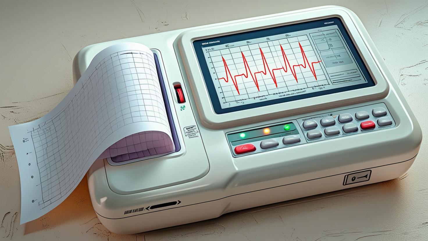
The electrocardiogram (ECG or EKG) machine, pioneered by Willem Einthoven in 1903, records the heart's electrical activity to detect conditions like arrhythmias or heart attacks. Early ECGs used heavy string galvanometers, but modern machines, like those from GE Healthcare, are compact, digital, and can transmit data wirelessly for instant analysis. Some portable ECGs, small enough to fit in a pocket, allow patients to monitor their heart at home, revolutionizing cardiac care with real-time diagnostics.
The 13 sections, in brief
Short summaries of each section. The full lessons, images and quizzes are in the Platform.
Overview
A man of fifty-two walks into an emergency department at ten past two in the morning with chest pain. A nurse takes him straight to a cubicle, attaches ten stickers to his chest and limbs, and presses a button. Ten seconds later a piece of paper comes out of a machine, and everything that happens to him for the next twenty-four hours is decided by what is on it.
Operations
Follow the signal from the cell to the paper. It starts with the electrodes. Ten of them: one on each limb, six across the chest. Each is a silver and silver chloride interface in a conductive gel, converting ionic current in the body into electron current in a wire.
Importance
The first argument is about a single decision and it is the most consequential one this device makes. The ECG determines who goes immediately for emergency reperfusion. A complete blockage of a coronary artery produces a characteristic pattern of ST elevation.
Physics
Concept 1, The Core Principle of Electrocardiography: Capturing the Electrical Symphony of the Heart Electrocardiography (ECG) is the science, and art, of transforming invisible ionic currents within the human heart into visible, quantifiable waveforms that represent the rhythmic choreography of depolarization and repolarization.
Features
Start here, because this is where most errors in the device originate. One on each arm and each leg. The right leg electrode is not a measuring electrode at all: it is a driven reference that actively cancels interference the patient is picking up. The limb electrodes see the heart in the frontal plane, from above, below and either side.
Applications
Every use of this device is one of four things. Deciding whether to activate an emergency pathway. Naming a rhythm. Detecting a systemic problem showing itself in the heart. Or clearing somebody for something. The fourth one is the largest by volume and the weakest by evidence.
Physiology
How a single cell produces a voltage, how millions of them sum into a field, how that field reaches the skin, and why damage to the muscle changes the shape of what arrives. Start at the cell, because everything else is a sum of this. A heart muscle cell maintains a large potassium gradient across its membrane, with the inside negative relative to the outside.
Safety
The device itself is almost incapable of injuring anybody. All of the harm in this device comes from what is done with the tracing. A missed infarction, a misread rhythm, a normal recording used to send somebody home, an artefact treated as disease, a lead reversal interpreted as pathology, or an automated interpretation accepted without looking.
Advancements
Forget release years. This device has a distinctive shape: the measurement was perfected early and has barely changed, and everything since has been about where the recording happens and who reads it. Somebody measured the heart's electrical activity from the surface of the body using an instrument sensitive enough to detect a millivolt, which required immersing the patient's limbs in buckets of salt solution.
In Research
A century old device with an unmeasured error rate The ECG is one of the oldest and most widely performed tests in medicine. And almost no hospital knows how often its own staff misread one. Studies that have measured it find error rates on clinically important findings that would be considered unacceptable in any other diagnostic process, including on tracings showing complete coronary occlusion.
Logistics
An engineer who understands this device is useful. An engineer who understands that this is the cheapest genuinely high-value diagnostic device in the library and its value is entirely contingent on somebody being able to read the output understands what is actually being sold.
Alternatives
An alternative is not a competing product. It is another way of answering the clinical question, and for this device the honest position is unusual. For the questions it answers, almost nothing competes. Its limitations are mostly answered by other tests used alongside it, not instead of it.
Ethical
This device produces a piece of paper that carries enormous authority. A printed tracing with a printed diagnosis looks objective in a way that a clinical impression does not, and it is treated accordingly. It goes in the notes, it is quoted in handovers, it appears in coroners' reports, and it is cited as evidence that a proper assessment was made.
Previous: EEG (Electroencephalogram) Machine. Next: Electrophysiology Mapping System. All 69 devices.
Learn it properly, inside the Platform
22 courses, the 69-device library with 13 sections per device, BMEssentials and BMEssence guides and MedTech AI. Start with a 7-day free trial, no card.
Start the 7-day free trial