
BMEssentia Platform / Device library / Electrophysiology Mapping System
Device library
Electrophysiology Mapping System
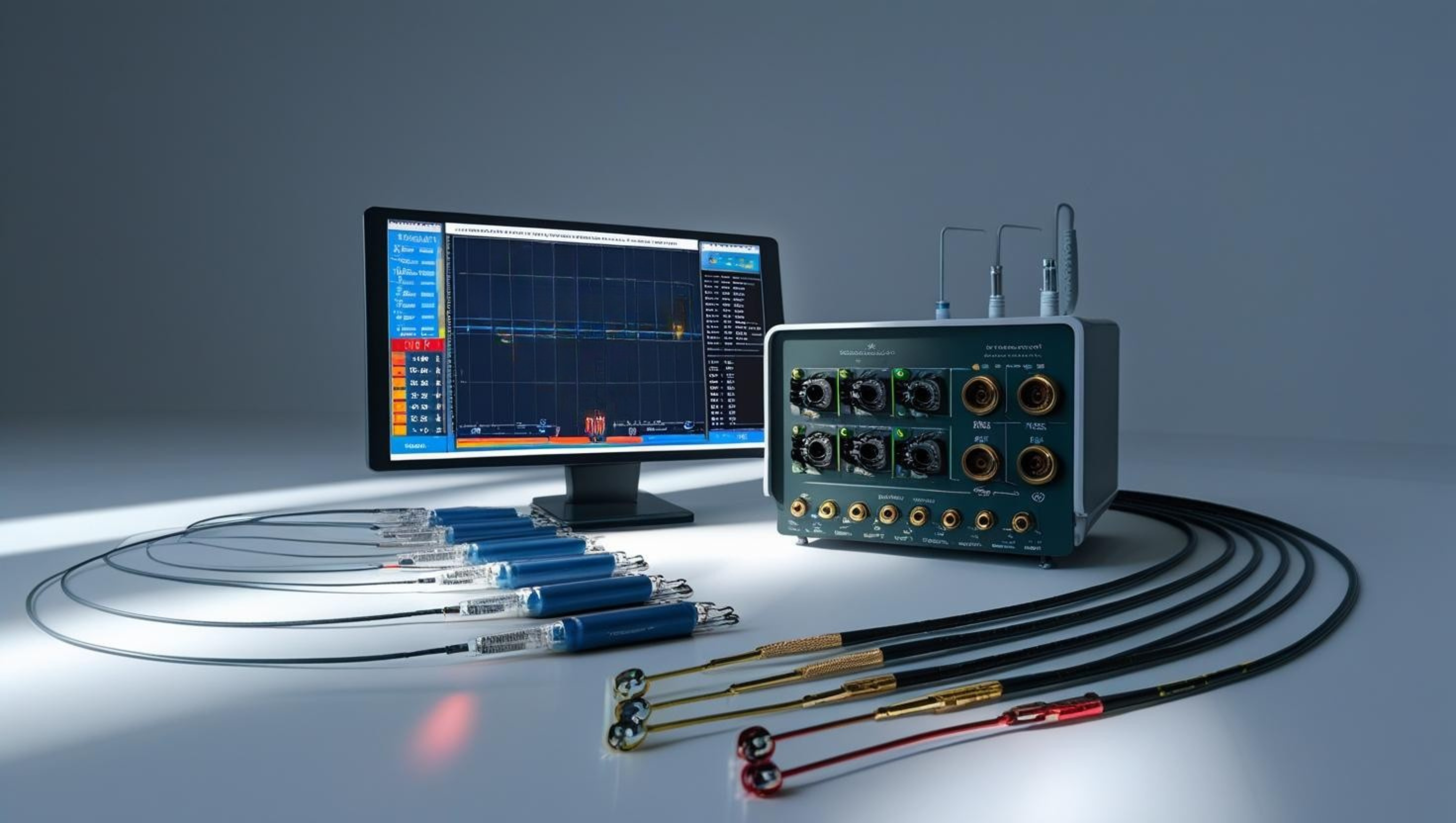
The electrophysiology mapping system is a sophisticated cardiac diagnostic tool that creates 3D maps of the heart's electrical activity to identify arrhythmia sources. These systems use catheter-based sensors and advanced algorithms to visualize electrical propagation patterns with millisecond precision. Modern platforms integrate real-time data acquisition with high-resolution anatomical mapping, enabling precise localization of abnormal pathways in conditions like atrial fibrillation. Some cutting-edge systems now incorporate artificial intelligence to predict optimal ablation sites and improve procedural outcomes. This technology has revolutionized treatment for complex cardiac.
The 13 sections, in brief
Short summaries of each section. The full lessons, images and quizzes are in the Platform.
Overview
A woman in her forties has been having episodes where her heart suddenly races to two hundred beats a minute for twenty minutes at a time, several times a year, for a decade. Drugs have not stopped it.
Operations
Access through a vein, usually in the groin, up into the heart. For left-sided work the catheter is passed from the right atrium across the wall between the two atria, through a deliberate puncture. Then two things happen at once, and understanding that they are separate is the key to the whole device.
Importance
The argument is cure. Most of medicine manages conditions. This one, in several arrhythmias, ends them permanently. A young person with a rhythm caused by an extra electrical connection between the atria and ventricles has a circuit that can be located and destroyed in a single procedure, with a success rate above ninety percent and essentially no recurrence.
Physics
Physics Involved in the Electrophysiology Mapping System: Concept 1, Principles of Electrophysiological Mapping and Cardiac Signal Acquisition An Electrophysiology (EP) Mapping System is the cardiac equivalent of a telescope for electricity, a real-time visualization platform that lets physicians see how electrical impulses travel through the myocardium, millisecond by millisecond.
Features
The foundation, because a timing measurement with no position attached is useless. Three weak magnetic field generators sit under the table, each producing a field that varies in strength across the space. A tiny sensor in the catheter tip measures how strong each field is at its location.
Applications
The extra connection that should not exist The clearest success in the whole field and the best place to start. Normally the top and bottom chambers of the heart are electrically insulated from each other, except at one point where the signal passes through deliberately and slowly.
Physiology
This device measures electricity in living heart muscle and then destroys some of it. Which means every signal on the screen is a statement about what cells are doing, and every ablation is a permanent change to how a wave will travel through that heart for the rest of the person's life.
Safety
Two categories of risk, and they arrive at different times. Things that go wrong during the procedure, which are mostly about catheters in a thin-walled chamber. And things that go wrong afterwards, which are caused by the ablation and which arrive after the patient has gone home.
Advancements
Six movements, and the shape is a specialty inventing itself. Catheters were put inside the heart to record electricity, which showed for the first time what the surface ECG had only implied. Somebody worked out that a circuit could be interrupted by destroying a few millimetres of tissue, which turned a diagnostic technique into a cure.
In Research
This specialty cures several arrhythmias outright, which is rare in medicine. And its largest procedure volume is for a condition it cannot map. Atrial fibrillation is treated by burning a ring around the pulmonary veins, which is an anatomical target chosen because triggers usually come from there. Not because the mapping identified the mechanism in that patient.
Logistics
A capital system that is essentially given away, and catheters that cost a great deal and are used once. And the lock-in here is stronger than anywhere else in this library. The mapping system only works with that manufacturer's sensor-equipped catheters.
Alternatives
The goal is not to map a heart. It is for somebody to stop having episodes that frighten them, or to stop being at risk from a rhythm that could kill them. Which means the honest comparison is nearly always against a tablet.
Ethical
Most procedures in this library treat something dangerous. A young person with episodes of racing heart is frightened and inconvenienced, and in most cases not in danger. They are being offered a several-hour invasive procedure, with a small risk of stroke, perforation and permanent pacemaker dependence, to treat something that was never going to kill them.
Previous: Electrocardiogram (ECG or EKG) Machine. Next: Electrosurgical Unit. All 69 devices.
Learn it properly, inside the Platform
22 courses, the 69-device library with 13 sections per device, BMEssentials and BMEssence guides and MedTech AI. Start with a 7-day free trial, no card.
Start the 7-day free trial