
BMEssentia Platform / Device library / EMG (Electromyography) Machine
Device library
EMG (Electromyography) Machine
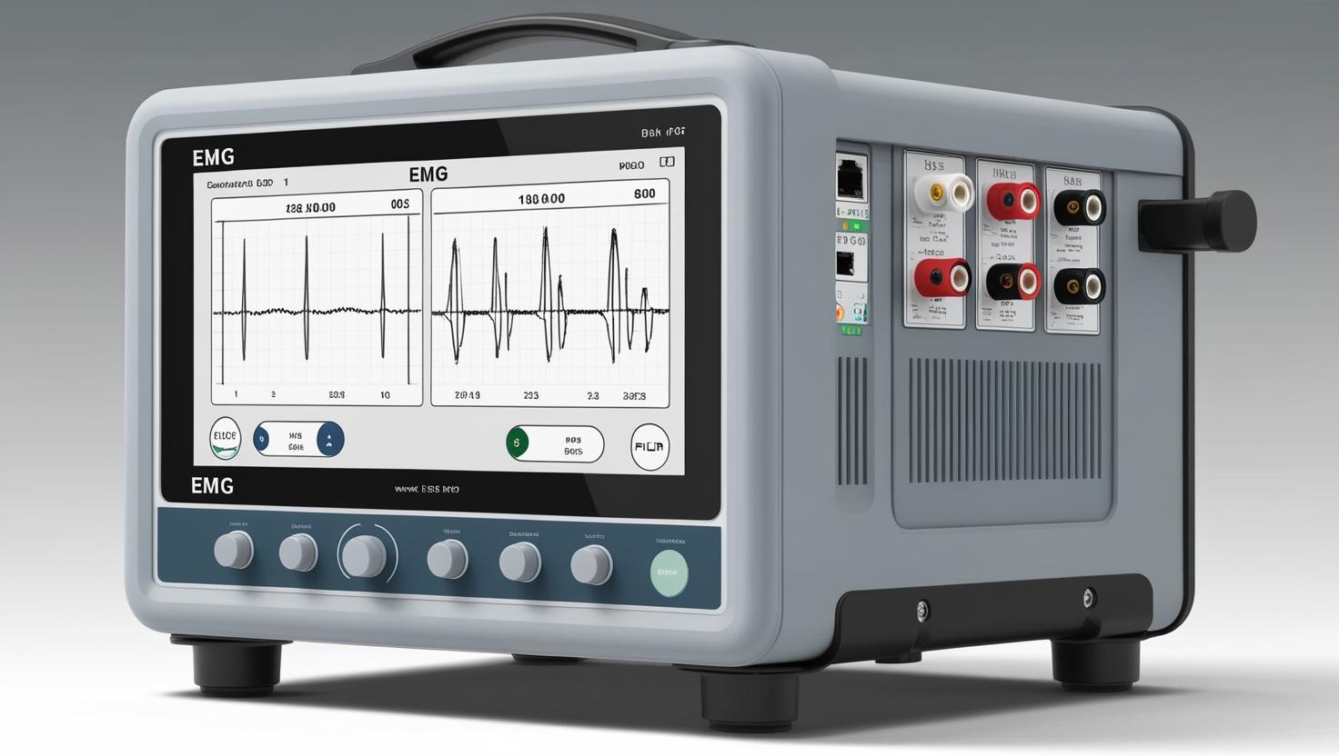
Electromyography (EMG) machines, developed in the 1940s, measure electrical activity in muscles to diagnose conditions like muscular dystrophy or nerve disorders. By inserting tiny needles or using surface electrodes, they detect signals as weak as a few microvolts. Modern EMG systems provide real-time data visualization and can integrate with nerve conduction studies for comprehensive neuromuscular assessment. Portable EMG devices now allow testing in outpatient settings, improving early diagnosis of conditions like carpal tunnel syndrome.
The 13 sections, in brief
Short summaries of each section. The full lessons, images and quizzes are in the Platform.
Overview
A woman of fifty-four has had weakness in her right hand for eight months. Her fingers are thinner than they were. She drops things. Two doctors have told her it is probably a trapped nerve in her neck and that it will settle. A needle is inserted into the muscle at the base of her thumb. There is no anaesthetic, because anaesthetic would abolish the very signal being measured. She is asked to relax completely.
Operations
Follow the two halves separately, because they are separate examinations. Nerve conduction studies start with stimulation. A surface electrode delivers a brief electrical pulse over a nerve, strong enough to depolarise every axon in it. Then recording. Surface electrodes over the muscle that nerve supplies, or over the nerve itself further along.
Importance
The first argument is about a question no imaging test can answer. When somebody is weak, the problem can be in five different places, and the treatment and prognosis differ completely for each. The motor neuron in the spinal cord. The nerve root. The peripheral nerve. The neuromuscular junction. Or the muscle itself.
Physics
Concept 1, Electrical Activation and Signal Genesis in Skeletal Muscle Electromyography (EMG) is the precise measurement of the electrical essence of muscle activity, the translation of ionic movement inside excitable tissue into an observable electrical waveform. The EMG device is not just a detector; it is a translator between biological excitation and electronic interpretation.
Features
The half of the device that puts energy in. A brief pulse, typically a fraction of a millisecond, delivered as a constant current so that the amount reaching the nerve does not change if skin resistance changes. Delivered through a handheld probe with two electrodes, positioned over the nerve.
Applications
Every use of this device answers one question in four different ways. Where is the lesion? In the motor neuron, the root, the nerve, the junction, or the muscle? And then three follow-ups: what kind of damage, how severe, and how long has it been there.
Physiology
How a nerve conducts, why myelin makes it fast, what a motor unit is, what happens when a nerve dies, and why muscle repairs itself the way it does. The foundation, because every measurement in the device is a property of this process. An axon maintains a negative interior. When depolarised past threshold, sodium channels open, sodium rushes in, and the interior briefly becomes positive.
Safety
This device carries a small set of real physical risks and one large one that is not usually counted as a risk at all. The pain is the safety issue. Not because pain itself is the harm, but because it determines whether the study is completed, and an incomplete study in a patient with motor neuron disease or an immune neuropathy is a delayed diagnosis with real consequences.
Advancements
Forget release years. This device has an unusual shape: the core measurement was established early and has hardly changed, and the meaningful advances have been in what the examiner can see while doing it and what else can be combined with it.
In Research
A test that is designed while it is performed Almost every diagnostic test in this library follows a protocol. This one does not. Which means the largest determinant of whether an EMG study answers the clinical question is a series of decisions made by one person in real time, and almost nothing is known about how good those decisions are. Nobody measures how often the muscles examined were the right ones.
Logistics
An engineer who understands this device is useful. An engineer who understands that this is a modest machine that consumes an hour of a rare specialist's time per patient understands what is actually being sold. An EMG system is one of the cheaper diagnostic devices in this library.
Alternatives
An alternative is not a competing product. It is another way of answering the clinical question, and here the question is always the same. Where in the chain from motor neuron to muscle fibre is the problem, and what kind of problem is it? Almost everything in this section answers part of that and nothing answers all of it.
Ethical
This is the only routine diagnostic test in this library that requires the examiner to hurt the patient repeatedly for an hour, with no anaesthetic, while the patient cooperates actively throughout. And the examiner frequently knows the diagnosis before the study is finished, while the patient lies there making conversation.
Previous: Electrosurgical Unit. Next: Endoscopes. All 69 devices.
Learn it properly, inside the Platform
22 courses, the 69-device library with 13 sections per device, BMEssentials and BMEssence guides and MedTech AI. Start with a 7-day free trial, no card.
Start the 7-day free trial