
BMEssentia Platform / Device library / Ophthalmoscope
Device library
Ophthalmoscope
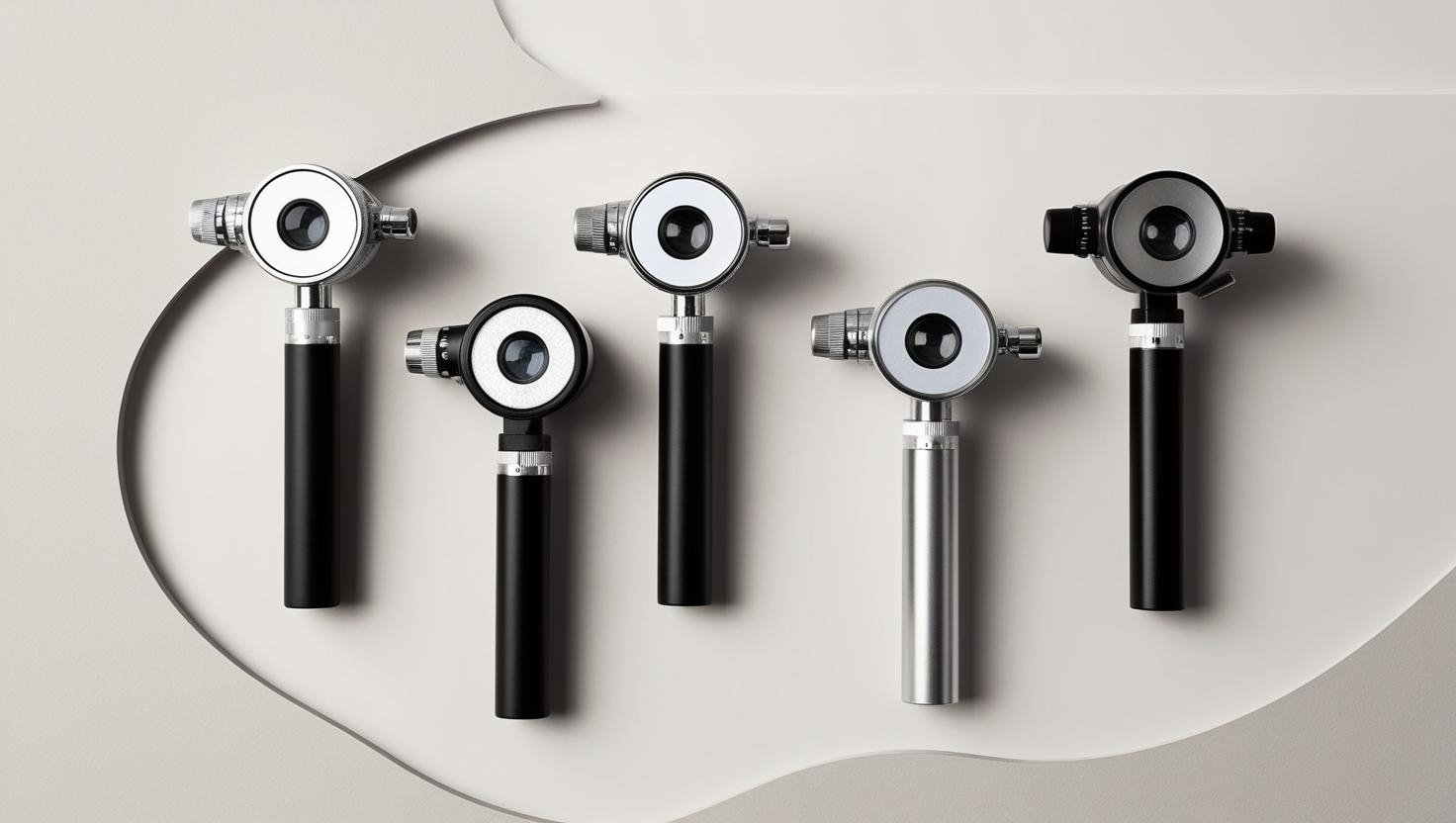
The ophthalmoscope, invented in 1851 by Hermann von Helmholtz, was the first device to allow doctors to see inside a living human eye, revolutionizing eye care. Originally a simple lens and mirror setup, it helped diagnose conditions like glaucoma and diabetic retinopathy. Modern ophthalmoscopes use advanced optics and LED lights for clearer retinal images, and some even capture digital photos for telemedicine. This tool remains essential for eye exams, aiding millions in preserving vision.
The 13 sections, in brief
Short summaries of each section. The full lessons, images and quizzes are in the Platform.
Overview
Alternate Names: Direct Ophthalmoscope, Indirect Ophthalmoscope, Funduscope Ophthalmoscope - A diagnostic tool for examining the internal structures of the eye, particularly the retina, optic disc, and blood vessels. Direct Ophthalmoscope - A handheld device providing a magnified (up to 15x) upright image of the retina, suitable for close-up viewing.
Operations
The ophthalmoscope operates on the basic principle of directing a beam of light through the pupil to illuminate the retina and its surrounding structures. The returning light, reflected from the retina, is then magnified by a series of lenses, allowing the examiner to view the back of the eye in detail.
Importance
The ophthalmoscope is indispensable in clinical practice because it allows for direct visualization of the retina, optic disc, and other internal structures of the eye, making it critical in diagnosing a range of eye and systemic conditions. Without ophthalmoscopic examinations, many conditions would remain undetected until they progress to more severe stages.
Physics
Physics Involved in the Ophthalmoscope Device: Concept 1, Optical Design and Retinal Visualization in the Ophthalmoscope The ophthalmoscope is a small handheld instrument, but inside its body lives an entire optical system built to do something that once seemed impossible: let a human being see through another person's eye and examine the living retina while light is still passing through it.
Features
Ophthalmoscopes are equipped with numerous features that make them versatile and essential for different diagnostic purposes. The device's design allows it to be used across various medical fields, offering insights not just into ocular health but also into systemic diseases.
Applications
Working Premise and Procedure (Step-by-Step) The ophthalmoscope allows healthcare professionals to visually inspect the internal structures of the eye, primarily focusing on the retina, optic disc, and blood vessels.
Physiology
The retina is a light-sensitive layer located at the back of the eye, responsible for converting light into electrical signals that the brain interprets as vision. It contains photoreceptors (rods for low-light vision and cones for color and high-acuity vision) as well as a complex network of cells and tissues essential for processing visual information.
Safety
Using bright light from the ophthalmoscope can cause temporary discomfort or irritation to the patient, especially if the eyes are sensitive to light. This can be mitigated by adjusting the light intensity or using dilating drops to make the examination easier.
Advancements
Recent advances in digital ophthalmoscopes allow for real-time imaging and recording of the retina. These systems store images digitally for future reference, aiding in tracking disease progression. This technology also allows for telemedicine, where retinal images can be shared with specialists remotely for second opinions or in cases where in-person examinations are not feasible.
In Research
The Device in Research and Specialized Applications Ophthalmoscopes are widely used in clinical trials and research focused on diabetic retinopathy, a leading cause of blindness. Researchers are developing AI-based ophthalmoscopy tools to screen for early signs of diabetic retinopathy, which would allow for early intervention and potentially prevent vision loss.
Logistics
Ophthalmoscopes vary in price depending on the type (direct or indirect) and the level of technology integrated into them. Basic direct ophthalmoscopes range from $200 to $800, making them accessible for most primary care settings. In contrast, indirect ophthalmoscopes are more expensive, ranging from $1,500 to $5,000 due to their advanced optics and wider field of view.
Alternatives
Fundus photography is a non-invasive method that uses a camera to capture detailed images of the retina, optic disc, and blood vessels. Unlike ophthalmoscopy, it provides a permanent visual record that can be referred to later. However, it lacks real-time flexibility of manual ophthalmoscopy and is often more expensive and time-consuming.
Ethical
Ethical Considerations and Future Directions Ethical Considerations in Retinal Screening: With the increasing use of ophthalmoscopes for screening purposes, especially in mass diabetic retinopathy programs and glaucoma detection, questions around patient consent, privacy, and data handling become important.
Previous: Nebulizer. Next: Orthopedic Implants. All 69 devices.
Learn it properly, inside the Platform
22 courses, the 69-device library with 13 sections per device, BMEssentials and BMEssence guides and MedTech AI. Start with a 7-day free trial, no card.
Start the 7-day free trial