
BMEssentia Platform / Device library / Retinal Laser Photocoagulation System
Device library
Retinal Laser Photocoagulation System
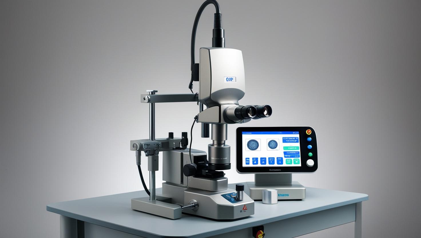
Retinal laser photocoagulation, pioneered in the 1950s by Meyer-Schwickerath, uses precise laser beams to treat conditions like diabetic retinopathy and retinal tears by creating tiny scars that seal leaking blood vessels or reattach the retina. Early systems used xenon-arc lamps, but modern devices, like the PASCAL laser, employ 532-nm green lasers for faster, less painful treatments with pattern technology. These systems can prevent severe vision loss in conditions like proliferative diabetic retinopathy, though they may cause minor side effects like temporary blurriness
The 13 sections, in brief
Short summaries of each section. The full lessons, images and quizzes are in the Platform.
Overview
A man of forty-one is sitting at a machine with his chin on a rest, and the ophthalmologist is about to deliberately destroy part of his retina. Not by accident. Not as a side effect. The destruction is the treatment. He has diabetes, and the blood supply to large areas of his peripheral retina has failed.
Operations
Follow the light, then follow the heat, because they are separate stories. It starts in the laser source. A solid-state crystal pumped by diodes, producing light at a wavelength chosen for how it interacts with the tissue. Then the delivery path.
Importance
The first argument is the strongest in ophthalmology and it is worth stating plainly. This device is the reason proliferative diabetic retinopathy is no longer a guaranteed cause of blindness. Before it, a patient whose eye began growing abnormal vessels was on a path that ended in a vitreous haemorrhage, a tractional detachment, and blindness. There was nothing to offer.
Physics
Physics Involved in the Retinal Laser Photocoagulation System: Concept 1, Optical Energy Delivery and Tissue Coagulation Control A retinal photocoagulator converts light into localized heat, sealing microscopic retinal leaks or abnormal blood vessels without cutting tissue.
Features
Start with the mechanism, because it is different from every other laser in this library. A surgical cutting laser vaporises tissue. A lithotripter fractures a stone. This one raises the temperature of a small volume of tissue until the proteins in it unfold and the cells die.
Applications
The application that built the device and the one that saved the most sight. Years of high blood sugar damage the smallest vessels. Areas of retina lose their blood supply. Starving retina releases a chemical signal demanding new vessels. The eye grows them, and they are useless: fragile, leaky, and growing forward into the vitreous rather than staying in the retina.
Physiology
Why the retina is so hungry for oxygen. What starving retina does about it. Why burning tissue fixes that. And what the burning costs. The most metabolically demanding tissue in the body Start here, because it explains why this disease exists at all. They run a continuous current even in darkness, pumping ions constantly to maintain it, and light reduces that current rather than creating one.
Safety
This device is unusual in the library because the harm is intentional. Every burn destroys retina permanently, and that is the treatment working correctly. Which means safety here is not about preventing damage. It is about keeping the damage exactly where it was meant to be, in the amount that was meant, and stopping it from reaching two places: the centre of the patient's vision, and anybody else's eye in the room.
Advancements
Forget release years. This device has an unusual shape: it solved a major cause of blindness, then spent decades being refined, and is now being displaced by a drug for much of what it treats. Somebody burned a retina with focused sunlight and observed that it scarred and stuck down. Crude, uncontrollable, and it established the principle.
In Research
A doctor sets a power on a console, fires a test shot, looks at how white the tissue went, and adjusts by eye. Which means the most widely used sight-saving treatment in the history of ophthalmology has no dosimetry. Nothing measures the energy arriving at the retina. Nothing accounts for pigmentation, for media clarity, for the lens in use, or for the calibration state of the machine.
Logistics
An engineer who understands this device is useful. An engineer who understands that this is one of the very few machines in the library with no consumable at all understands what makes it different from everything around it. There is no pack, no probe, no cartridge, no per-case cost. You buy the machine and a few lenses and you treat patients for fifteen years.
Alternatives
An alternative is not a competing product. It is another way of answering the clinical question, and here the question depends on what the laser is being used for. To weld a retinal tear, the alternatives are few. To stop abnormal vessel growth, there is now a genuine competitor that works better.
Ethical
Almost every device in this library tries to avoid harming the patient. This one harms them deliberately, and that is the treatment. You are destroying functioning retina, permanently, in a person who can currently see, to prevent something worse later. Which makes the consent conversation unlike any other in ophthalmology. The patient is not accepting a risk.
Previous: Pulse Oximeter. Next: SPECT Scanner. All 69 devices.
Learn it properly, inside the Platform
22 courses, the 69-device library with 13 sections per device, BMEssentials and BMEssence guides and MedTech AI. Start with a 7-day free trial, no card.
Start the 7-day free trial