
BMEssentia Platform / Device library / SPECT Scanner
Device library
SPECT Scanner
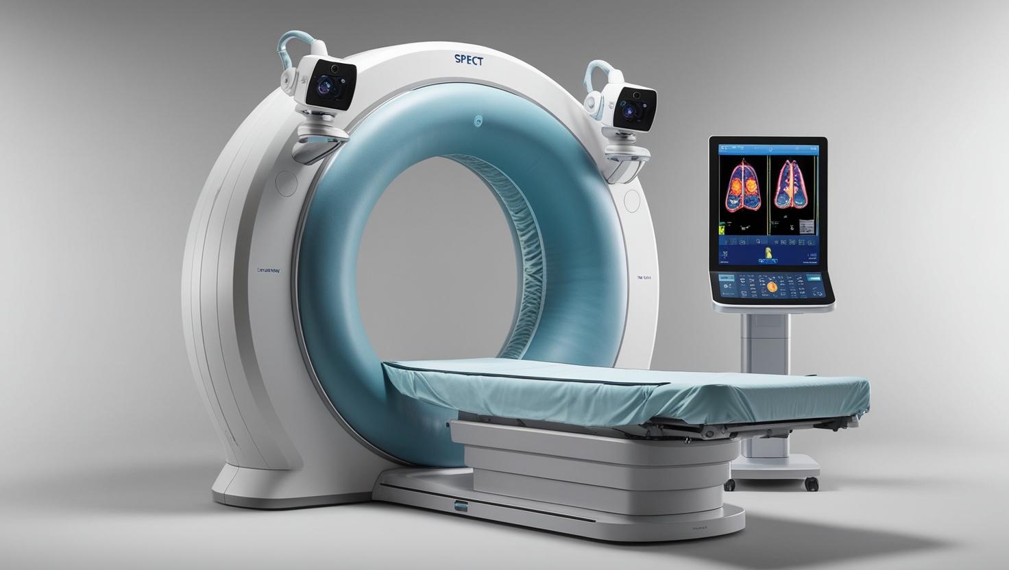
A SPECT (Single-Photon Emission Computed Tomography) scanner is a nuclear imaging device that creates 3D functional images of organs by detecting gamma rays emitted from radioactive tracers. This advanced diagnostic tool combines a gamma camera with rotational capabilities to capture multiple cross-sectional views of metabolic activity. Modern SPECT systems often integrate with CT scanners (SPECT/CT) to overlay functional and anatomical images for precise localization of abnormalities. Clinically essential for cardiology, neurology, and oncology, it evaluates blood flow, brain function, and tumor activity with superior sensitivity than conventional imaging. New detector materials and.
The 13 sections, in brief
Short summaries of each section. The full lessons, images and quizzes are in the Platform.
Overview
A woman in her sixties is walking on a treadmill in a hospital basement while a nurse watches a monitor and a cardiologist watches her face. She has been having chest pain when she climbs stairs, and nobody can tell her whether that is her heart or her anxiety, because her resting ECG is normal and nobody has ever looked at her arteries. The treadmill speeds up. Her heart rate climbs.
Operations
Follow the whole chain and the machine explains itself. A tracer is prepared, which for most clinical SPECT means combining a radioactive isotope with a biologically active compound in a small vial, frequently in the hospital's own radiopharmacy on the morning it is used.
Importance
The case for SPECT rests on one thing that sounds modest and is not: it is the widely available, affordable, logistically forgiving way to image a biological process.
Physics
1. Every SPECT image begins in the nucleus. Deep inside an atom lies the birthplace of the signal, an unstable core seeking balance. This atomic instability is not random chaos but a physical drive toward equilibrium, expressed through the elegant process of radioactive decay.
Features
The collimator, and the machine that throws away its own signal Here is the problem the collimator exists to solve, and it is worth feeling it before seeing the answer. A radioactive atom inside the patient decays and emits a single gamma photon. That photon flies off in a completely random direction. It might travel toward the detector. It might travel toward the floor.
Applications
Cardiac perfusion, which is the largest single use of this machine Start with the clinical problem, because the elegance of the answer only lands once you feel the difficulty. A patient has chest pain on exertion. The question is whether a coronary artery is narrowed enough to matter.
Physiology
Every image this machine produces is a statement about a physiological process. which means the physiology is not background to the device. It is the device. A CT image can be interpreted, imperfectly, by someone who knows anatomy. A SPECT image cannot be interpreted at all by someone who does not know what the tracer does in the body, because the image is a map of a process rather than a map of a structure.
Safety
This machine inverts the arrangement that every other imaging device follows. In an X-ray or CT room the machine produces radiation, the patient absorbs some, and switching the machine off makes the room safe. Here, the source is injected into the patient.
Advancements
Forget release years. The shape is what predicts direction and it stays true for a decade. This modality moved in five waves, and one of them is unusual: a long period where almost nothing changed. The camera was invented and then barely altered for decades. A large sodium iodide crystal, an array of photomultiplier tubes, and position computed from the weighting of their signals.
In Research
SPECT is the most widely deployed functional imaging modality on earth. There are many times more gamma cameras than PET scanners. It has been in clinical use for decades, its physics is completely understood, and its core applications are among the most performed studies in nuclear medicine.
Logistics
An engineer who understands how a gamma camera works is useful. An engineer who understands how a generator is delivered every Monday, how kits clear customs as pharmaceuticals rather than as devices, and what a department does in the week a reactor goes offline, runs a nuclear medicine service or a distributorship.
Alternatives
An alternative is not a competing product. It is another way of answering the clinical question, and the honest comparison changes completely depending on what is being asked, about whom, and in what circumstances. And this section is unusual, because SPECT is the modality under the most genuine competitive pressure of any device in this library.
Ethical
This modality raises ethical questions that most imaging does not, for four specific reasons. The radiation source is injected rather than external, which puts it beyond recall the instant the plunger moves. Its supply depends on a handful of ageing reactors, which means allocation decisions during shortages are made globally and affect patients who will never know why their study was cancelled.
Previous: Retinal Laser Photocoagulation System. Next: Spirometer. All 69 devices.
Learn it properly, inside the Platform
22 courses, the 69-device library with 13 sections per device, BMEssentials and BMEssence guides and MedTech AI. Start with a 7-day free trial, no card.
Start the 7-day free trial