
BMEssentia Platform / Device library / Transesophageal Echocardiography (TEE) Probe
Device library
Transesophageal Echocardiography (TEE) Probe
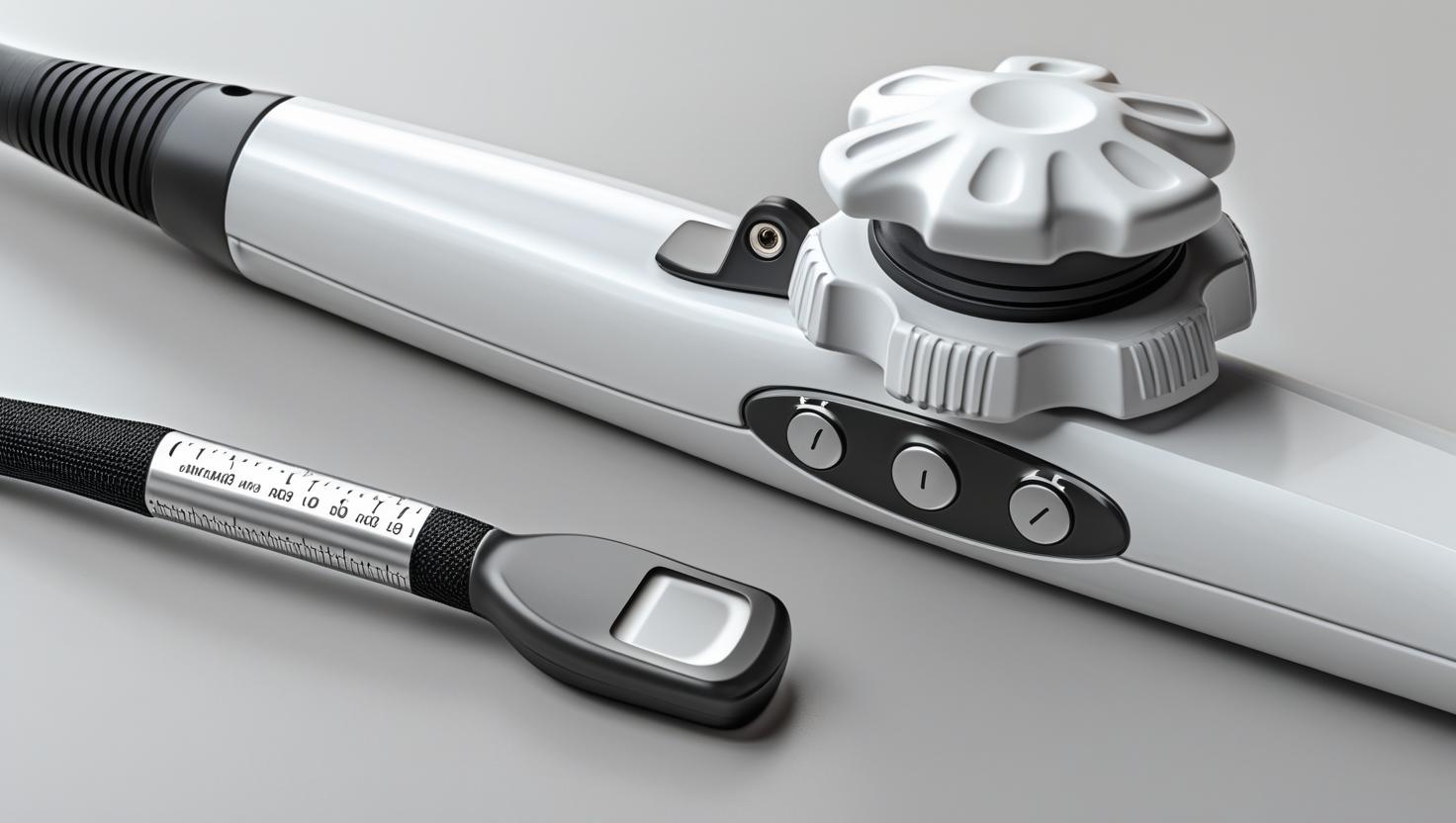
The Transesophageal Echocardiography (TEE) probe provides detailed images of the heart by being gently inserted into the esophagus, which lies close to the heart. Because of its proximity, it offers clearer and more accurate visuals than standard external ultrasounds. TEE is especially valuable during heart surgeries or when detecting blood clots, valve problems, or infections. The procedure is safe and usually performed under mild sedation for patient comfort.
The 13 sections, in brief
Short summaries of each section. The full lessons, images and quizzes are in the Platform.
Overview
A patient is on a table with their chest open and their heart stopped, and the surgeon has just finished repairing a mitral valve. Everybody wants to know whether the repair worked. There is no way to test it: the heart is not beating, the chamber is empty, the valve is being held by sutures that have not yet been asked to do anything.
Operations
Follow the probe and the device explains itself. It is a flexible shaft, roughly the diameter of a finger, with a transducer array at the tip and a handle carrying two control wheels. Those wheels flex the tip up and down and side to side by pulling on internal cables, exactly the mechanism described in the surgical robot device, and with the same silent failure mode when a cable stretches.
Importance
The case for this device rests on three things it does that nothing else can, and the first is the strongest. Intraoperative assessment during cardiac surgery changes the operation while it is still happening. A valve repair is assessed under load, immediately, and a repair that leaks is revised in the same anaesthetic rather than in a second operation months later.
Physics
1, Functional Architecture and Working Principle The TEE probe is a specialized ultrasound transducer designed to capture exquisite, real-time images of the heart from inside the esophagus, just millimeters behind the left atrium. By moving the imaging source inside the body, TEE bypasses the interference of ribs, lungs, and chest wall, revealing cardiac anatomy with extraordinary clarity and dynamic detail.
Features
Why the oesophagus, and why frequency is the answer Start with the problem the device solves, because everything else follows. Higher frequency gives finer resolution, because the wavelength is shorter and two structures closer together can still be distinguished.
Applications
Not a diagnostic test here. A continuous monitor that changes the operation while it is happening. The pre-bypass study confirms the diagnosis and frequently modifies the plan. The mechanism of a valve problem seen at this resolution is not always what the pre-operative study suggested, and the repair strategy depends entirely on mechanism.
Physiology
This section runs in two directions and they are unusually separate. What the probe does to the oesophagus, which is a mechanical story about a tube that was not designed to hold an instrument. And what the probe reveals about the heart, which is where the entire clinical value sits.
Safety
The complication rate for this device is low. Genuinely low, by any standard. Which produces the specific hazard worth naming first. A procedure that goes well thousands of times in a row stops feeling like a procedure. The probe becomes routine, the checks get compressed, and the rare complication arrives in a department that has not thought about it in two years.
Advancements
Forget release years. The shape predicts direction, and this device has a shape driven almost entirely by one number: how many elements can be fitted into a tip small enough to swallow. Somebody put a transducer on a swallowed cable. Single element, no image in any modern sense, and it proved the acoustic argument: the view from behind the heart is fundamentally better.
In Research
It is standard in cardiac surgery. It changes operations in progress. It has been used for decades. And almost nobody can tell you whether it improves outcomes. Not because the evidence is negative.
Logistics
An engineer who understands this device is useful. An engineer who understands that the probe costs a large fraction of the machine it plugs into, is destroyed by mishandling, and cannot be used without a disinfection facility nobody costed understands what is actually being sold.
Alternatives
An alternative is not a competing product. It is another way of answering the clinical question, and the question here is almost never "image the heart." It is far more specific than that. Is there clot in the appendage. Did this repair work. Is this valve infected. Why is this patient shocked.
Ethical
Most devices in this library raise ethical questions about what is done to a patient. This one raises a narrower and sharper question, and it is worth stating exactly.
Previous: Transcutaneous Electrical Nerve Stimulator (TENS). Next: Ultrasound (SONAR) Machine. All 69 devices.
Learn it properly, inside the Platform
22 courses, the 69-device library with 13 sections per device, BMEssentials and BMEssence guides and MedTech AI. Start with a 7-day free trial, no card.
Start the 7-day free trial