
BMEssentia Platform / Device library / Ultrasound (SONAR) Machine
Device library
Ultrasound (SONAR) Machine
Ultrasound machines, first used medically in the 1950s, harness high-frequency sound waves to create real-time images of organs, fetuses, or blood flow, without radiation. Inspired by sonar technology from World War II, early devices produced basic 2D images, but modern systems, like those from GE Healthcare, offer 3D/4D imaging and Doppler for assessing blood flow. Portable ultrasound units, small enough to connect to smartphones, are now used in remote areas, transforming diagnostics in obstetrics, cardiology, and emergency medicine.
The 13 sections, in brief
Short summaries of each section. The full lessons, images and quizzes are in the Platform.
Overview
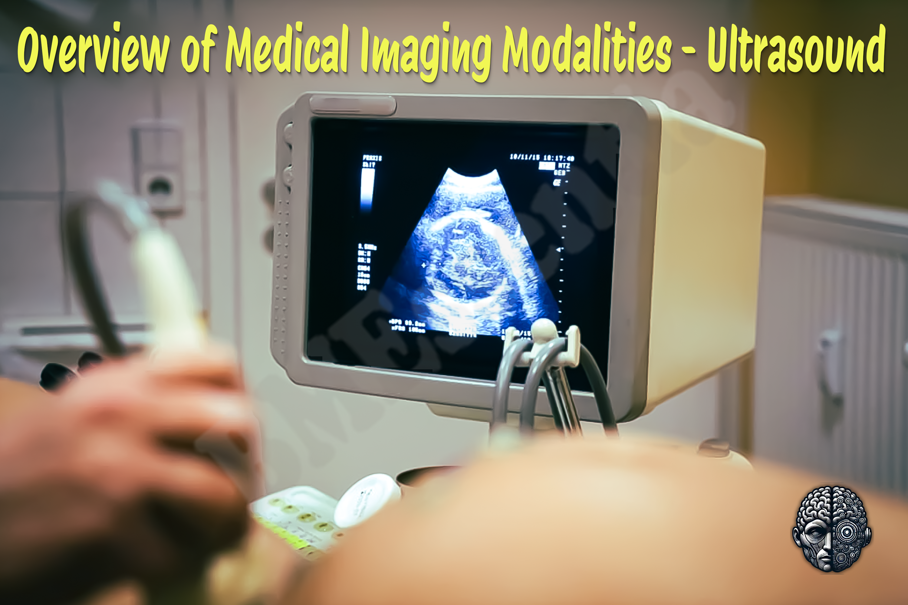
A woman is lying on a couch and a sonographer is pressing a probe into her abdomen. On the screen beside them, a grey and grainy picture moves in real time, and somewhere inside it is a four-chambered heart the size of a grape, beating at a hundred and fifty times a minute.
Operations
Follow the pulse, because the whole device is one pulse repeated a few million times a minute. It starts in the transducer. A layer of piezoelectric material, cut into a row of many small elements. Apply a voltage and the material deforms. Apply a short electrical pulse and the element flexes sharply, producing a brief burst of sound.
Importance
The first argument is about reach, and no other imaging device in this library comes close. Ultrasound is the only cross-sectional imaging modality that is genuinely available to most of the world's population. It needs no shielded room, no cooling, no three-phase power, no radiation licence and no helium.
Physics
Physics Involved in the Ultrasound Device: 1. Ultrasound imaging is built entirely on the behavior of mechanical sound waves traveling through biological tissue. Unlike electromagnetic waves (such as light or X-rays), sound needs a medium, molecules that can compress and expand to carry energy. In medical ultrasound, these are typically the molecules of soft tissue, fluid, or bone.
Features
The transducer, and why it is the whole machine Start here, because the console is largely a computer and the probe is the instrument. Certain materials deform when a voltage is applied across them, and generate a voltage when they are deformed. Which means the same element is both the speaker and the microphone.
Applications
The largest single application worldwide and the one most people have experienced. Confirming the pregnancy is inside the uterus, which is the question that matters most, because an ectopic pregnancy ruptures and kills. Dating it accurately, which determines when a baby is considered premature, when induction is offered, and how growth is judged for the rest of the pregnancy.
Physiology
What the body does to the sound: reflects it, absorbs it, scatters it, bends it, blocks it entirely. And what the sound does to the body: heats it, and in the right circumstances tears it. The property that creates every image in this device. Every tissue has a property combining its density with the speed of sound through it.
Safety
This device has a reputation for being harmless, and that reputation is the safety problem. There is no ionising radiation, which removes the one risk most clinicians and patients think about, and it produces a general belief that ultrasound is free. It is not free.
Advancements
Forget release years. This device has a distinctive shape: the physics was settled early, and almost every advance since has been about processing, portability and who is allowed to hold the probe. Sound was used to find things that could not be seen. Industrial flaw detection and underwater ranging established pulse-echo before anybody pointed it at a person.
In Research
Every other imaging device in this library can be audited. Somebody reviews the dataset and finds what was missed. Ultrasound cannot, because the images of what was missed do not exist. Which means nobody knows the miss rate for abdominal ultrasound, for obstetric anomaly scanning, or for point of care examinations.
Logistics
An engineer who understands this device is useful. An engineer who understands that the probes cost more than the machine over its lifetime understands what is actually being sold. A console lasts seven to ten years. Probes get dropped, cables get crushed by chair wheels, lenses wear, elements die, and an intracavity probe damaged by disinfectant is unrepairable.
Alternatives
An alternative is not a competing product. It is another way of answering the clinical question, and here the comparison turns on four things. Does the question need a complete survey or a focused look? Does it need motion? Does it need to be reproducible months later? And is there anybody available who can perform it?
Ethical
Most devices in this library raise ethical questions about how they are used on a patient. This one has been used, at population scale, to decide which children are born. Ultrasound made fetal sex determinable early, cheaply and non-invasively, and in societies with a strong preference for sons it enabled sex-selective abortion on a scale large enough to distort national sex ratios by millions.
Previous: Transesophageal Echocardiography (TEE) Probe. Next: Urodynamic Testing System. All 69 devices.
Learn it properly, inside the Platform
22 courses, the 69-device library with 13 sections per device, BMEssentials and BMEssence guides and MedTech AI. Start with a 7-day free trial, no card.
Start the 7-day free trial